Wednesday, March 16, 2011

I.C.A :4TH PART –INTRACRANIAL PART

Bismillah..I just suddenly want to establish the details here in this blog..and, the complete table draft you all can download by clicking here: >>...linking...<<    

CIRCULUS ARTERIOSUS of WILLIS 
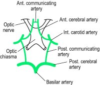


INTERNAL CAROTID ARTERY :4TH PART –INTRACRANIAL PART  


BR
(In Subarachnoid Space)

Course point

Area Supply


OPHTHALMIC ARTERY
Enter optic canal below optic nerve
Orbit

POST. COMMU. ARTERY
Communicating with Post. Cerebral Artery 


ANTERIOR CHOROIDAL ARTERY

·         Pass backward along optic tract around cerebral peduncle as far as LGB
·         Enter Inf horn of Lat Ventricle


-          Choroid plexus of Lat Ventricle
-          Optic tract
-          LGB
-          Internal Capsule;(post part of post limb,retrolentiform,sublentiform)

Terminal BR (Below Ant Perforated Subs.)


ANTERIOR CEREBRAL ARTERY
Reaching Median Longitudinal Fissure:
·         Pass forward & medially above optic chiasma

Reaching Medial surface of cerebral hemisphere:
·         Bend forward in the fissure & turn sharply upwards to reach the surface
·         Lies below the rostrum
·         Runs in callosal sulcus

End:
          ·         Above Splenium by turning upwards infront of parieto-occipital sulcus
1.       Cortical br:
-          Med.  surface of hemisphere (from frontal pole to parieto-occipital sulcus).
-          Upper 1’’ breadth of Lat. surface (as far as upper end of parieto-occipital sulcus).
-          Med. 1/2 of Orbital surface of frontal lobe.

2.       Central br:
-          Anterior part of corpus callosum
-          Anterior part of Ant. limb of Internal Capsule
-          Septal region (including SeptumLucidum)

3.       Callossal br:
              -          All corpus callosum EXCEPT                           splenium.


MIDDLE               CEREBRAL ARTERY

         ·        Pass Lat. in stem of lateral sulcus
         ·         Turn backwards & upwards in Post. Ramus of Lat. sulcus (crossing over Insula).


1.       Cortical br:
-          Lat. surface of cerebral hemisphere EXCEPT  ;
i)1 inch breadth  along Sup. border,
ii) Lat. surface of Occipital lobe.
iii) narrow strip on the temporal lobe close to Inf. border.

2.       Central br: (may include Chorcots Artery)
                   -          Corpus Striatum
                   -          Internal Capsule;(Post 1/2                           of Ant limb & Ant part of                           Post limb).   


Friday, December 24, 2010

Anatomy dan Student


'Antara Anatomy dan Study'

..Bismillahhirrahmaanirrahiim..

..Assalamualaikum/kunna..

Saya sangat pasti jika anda belajar anatomy, anda akan berhadapan dengan buku2 teks yang ber'volume'2...dan ini menjadi satu lumrah kebiasaan bagi mereka yang menceburi bidang MEDIC ini...sama lah juga bagi insan yang menaip di sini..~huahaha

Bak kata senior2, "I've been there."...dan ku akui agak susah untuk mengingati (bukan hafal erk) 'term2' MeDIC dalam BI, apatah lagi dalam Bahasa Latin!! Demikian itu, terdetik rasa di hati ini nak berkongsi tentang bagaimana diri ku ini mendalami ilmu Anatomy...

....dan tidak ku malu untuk mengatakan bahawa diri ku banyak mengambil cara2 dan teknik2 daripada orang lain bagi memperkemas sistem pembelajaran ku yang pada asalnya pun agak berterabur...maklum lah, diri ku masih dalam proses pematangan lagi ketika ini..~huhu

Kumpul punya kumpul, akhirnya ku temui jalan2 yang 'simple' dan paling asas sekali...


First Step : Visualization 

"Most people better learn anatomy by seeing how human body performs"


Kebenaran penyataan di atas telah terbukti berdasarkan pemerhatian terhadap rakan2 satu 'badge' ku...namun, lebih baik sekiranya seseorang itu memilih konsep pembelajarannya yang bersesuaian dengan personalitinya...

Namun demikian, ku cadangkan supaya belajar lebih kepada konsep gambaran, jujur ku katakan, kebanyakan pelajar kurang menyukai timbunan buku2 tanpa ada gambar2 untuk diperjelaskan kandungannya..soalnya sekarang, bagaimana??...Anda boleh belajar melalui buku2 ATLAS ataupun dengan software2 yang berlapis2 tersusun dalam rangkaian cyber zaman ini....ataupun, nak nampak 'advanced' skit, LUKIS SeNDIRI LAH..~huhu  


Namun, pendekatan kita dalam bidang MeDIC ini lagi manis rasanya jika digauli sekali dengan unsur2 hakikat Islam itu sendiri...contohnya dalam Embryology, bagaimana pada kaca mata kita sebagai seorang Muslim mempelajari proses2 kejadian manusia yang telah beratus ribu tahun terkandung dalam Al-Furqan??.... contoh video pembelajaran:


Second Step : Memorize In your Own Creativity 

"It is easier to memorize unfamiliar terms if you create certain rules"

'Rules' boleh ku tafsirkan kepada perkataan 'perATURan'...maksudnya, kaedah kita sendiri..dan ini bergantung kepada kreativiti individu masing2...

...contohnya seperti Peta Minda, lakar Jadual, menggariskan rangkaian2 vector dan etc dalam cubaan kita untuk memudahkan proses pembelajaran dan sekaligus mengingatinya dengan lancar dan sistematik...[ Latin + BI = my language ]...

....sistem ni pun Ok asalkan menepati cita rasa kita..dan ini sedikit sebanyak dapat memberi perangsang kepada kita untuk memperbanyak lagi bentuk2 'karya' kita untuk setiap topik yang berlainan...

"In another words, creating such rules and groups to memorize the terms can tremendously increase efficiency of your study of anatomy."..huhu, trust me...=D


Third Step : Take Self Tests 

"If repetitison is the mother of a skill then self test is father of the skill."


Oleh itu, amat bagus sekali pada mereka yang tidak memisahkan rangkaian ini :

STUDY ====>TEST

Semasa belajar, amat penting sekali bagi individu itu memeriksa setiap daripada langkah2 sistem pembelajarannya ....demikian itu, tidak hairan lah banyak kuiz2 serta latihan2 bagi mereka yang mempelajari Anatomy ini...khulasafnya, buatlah latih tubi walau sedikit...kerana Allah swt akan menilai setiap detik dan minit bagi hambaNya yang berusaha untuk kemajuan diri dan agama...

...Cukup setakat itu sahaja penulisan ku..sekadar 3 langkah yang mana telah menjadi asas dalam pembelajaran Anatomy berdasarkan pengalaman pengamal2 perubatan di sekitar dunia dan Mesir...

..Yang paling penting, BETULKAN NIAT dan perbanyakkan amalan2 kebaikan..InsyAllah..dipermudahkanNya... 

~wasalam~


4th Ventricle


Site : between = Ant : pons & upper part of medulla
Post : cerebellum

Shape : tent-shaped (side), diamond-shaped (behind)


Boundaries : Above & Lat = Sup cerebellar peduncle
                    Below & Lat = Inf cerebellar peduncle, Cuneate and Gracile tubercles

Angles : 
  • upper angle
  • lower angle 
  • Rt & Lt lateral angles


Floor : diamond-shaped
  • Upper pontine part : Facial colliculus, Median eminence, Pontine vestibular area, Sup fovea
  • Lower medullary part : Vagal trigone, Hypoglossal trigone, Vestibular area, Inf fovea  

Roof :

 a) Upper part => med border 2 sup cerebellar peduncles & sup medullary velum

 b) Lower part => 

         1) upper area : nodule of cerebellum & inf medullary velum on each side
    
        2) lower area : ependyma & pia mater, invaginated by choroid plexus
                                       
 *lowest part : F. of Magendi which open to subarachnoid space* 

 c) Recesses => Median recess, 2 Lat recesses ( open to subarachnoid space by F. of Luschka ) 


Connections : 
  • Upper angle : aqueduct of sylvius => connect 4th ventricle to 3rd ventricle
  • Lower angle : central canal of closed medulla => connect 4th ventricle to central canal of spinal cord
  • Roof : F. of Magendi & F. of Luschka => connect 4th ventricle to subarachnoid space    


Interpeduncular Fossa

Shape : rhomboidal-shaped space

Site : central region of the base of the brain

Boundaries : Ant = optic chiasma
                    Ant-Lat = optic tracts
                    Post-Lat = cerebral peduncle
                    Post = upper border of pons


Contents : 
  • Tuber cinereum
  • Infundibulum
  • 2 mamillary bodies
  • Post perforated substance
  • C3 nerve

Relations : 
  • Circle of willis (overlies the fossa)
  • 3rd ventricle (above it)
  • Pituitary gland (below it)
  • Ant perforated substance (anterolateral to it)


Tuesday, December 14, 2010

NeuroAnatomy (spinal cord)


Somebody missed the first Lecture?? give a little of your time to spare//


Friday, December 10, 2010

Simplying The Development of the FACE...Let's Revised

FACE DEVELOPMENT
From 5 processes/prominences:
a) 2 Maxillary process    ~~~~~~~>1st arch (mesoderm)
b) 2 Mandibular process ~~~~~~~>1st arch (mesoderm)
c) 1 Frontonasal process~~~~~~~~>proliferation of mesenchyme (buccopharyngeal)

STOMODEUM =  is a depression between the brain and the pericardium in an embryo, and is the precursor of the mouth and the anterior lobe of the pituitary gland.


(Cranially)
Frontonasal process 

Maxillary process      S    Maxillary process

2 Mandibular processes 
(caudally)

Diff. and Fusion..


FrontoNasal prominence :          

1.  Nasal placodes -----------> invaginates------------>FORM ===> Nasal pits, Lat  and Med nasal prominences

                                       grow medially

2.   Maxillary prominences --------------> compress Med nasal prominences ====> MERGE


Fate:                                                              

FrontoNasal prominence -------------> 
forehead, 

bridge of nose


Med nasal prominence ------------------> philtrum of upper lip, t
ip and crest of nose

Lat nasal prominences ----------------> ala of the nose


INTERMAXILLARY segment = free terminal part of frontalnasal prominence 
( lies between tips of 2 maxillary prominences)


Derivatives:


Labial component : philtrum of upper lip

Palatal component : triangular 1ry palate

Upper  jaw component : 4 incisors teeth


2 Maxillary prominences : 
  • Part of cheek upper lip (Except philtrum) 
  • upper jaw (Except median part carrying 4 incisors)
  •  2ry palate


2 Mandibular prominences

  • (fused) + Maxillary prominences ---------->most of the cheek
  • (fused) + Each other medially----------> lower lip & jaw 


click this link for the animation of Development of Face and Palate :
>>> animation <<<

::


: